Diagnosing and Treating Pet Fractures
Veterinarians describe fractures by which bone was affected, the fracture location within the bone, the shape or configuration of the break, and whether a foreign object or the bone itself punctured your dog’s skin.
If your dog was involved in an accident and has a suspected fracture, take your pet to the primary care veterinarian’s office or an emergency veterinary clinic right away. The doctor will check for internal injuries, which is crucial because some can be life-threatening.
Confine pets with fractures by encouraging them to lie in a small but proportionally sized place such as a box for smaller cats and dogs or a crate or kennel for larger dogs. Move them only to allow for urination or defecation or to maintain cleanliness. Call the primary vet or emergency clinic at once for guidance. Do not administer medication or attempt to perform treatment of any kind unless advised by a veterinarian.
Severity
Incomplete “greenstick” fractures may occur in young animals and can appear like a bend in the bone with cracks extending partway through the bone’s circumference.
Complete fractures are breaks that go from one side of the bone to the other, creating two or more bone fragments.
Complete fractures have subdivisions based on their morphology:
Transverse fractures are linear and break 90° perpendicular to the length of the bone.
Oblique and/or spiral fractures occur at an angle along the length of the bone, creating two sharply pointed bone fragments.
Comminuted fractures can be the most severe because the bone fractures in three or more pieces with varying sizes and shapes of fragments. Comminuted fractures can be associated with the most significant soft tissue swelling and instability.
Fractures and Whether the Skin Is Punctured
Bone breaks that puncture through the skin are called open fractures. Open fractures may also occur when objects forcibly enter the skin with sufficient force to damage bones. Open fractures may result in infection due to the bacteria entering the wound.
Closed fractures remain beneath the level of the skin.
Signs & Symptoms
Bones have innervation, which is why fractures are painful. Pets with fractures in the front or hind legs will hold up the affected limb, not wanting to bear weight due to pain. Depending on the number of fractures, their location, and their severity, some pets will be able to put minimal weight on the limb, while others may refuse to walk. Swelling or abnormal limb movement may be visible. Dogs in distress often lick or bite at the injured area, whimper, yelp, pant, or show signs of aggression such as biting or growling.
When trauma is involved, besides broken bones, internal organs may be damaged by:
- Contusions (bruising)
- Hemorrhage (bleeding)
- Laceration (cuts)
- Puncture
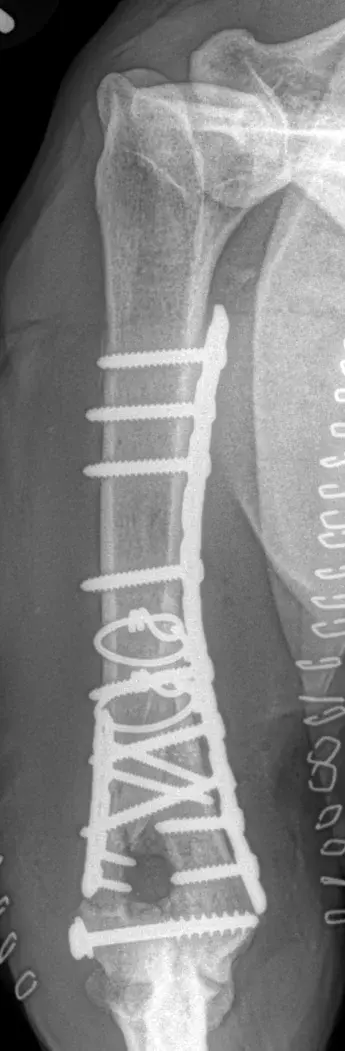
Multiple bone plates, screws and cerclage wires may be used to stabilize fractures. This common – and very complex – injury is often described as a “T” or “Y” fracture of the humerus and results in three separate segments including an intra-articular component. The joint surface is reconstructed first followed by application of bone plates both medially and laterally in neutralization.
Diagnosis
Veterinarians thoroughly examine pets, check for injuries to bones and internal organs, and order X-rays of the affected limb(s) and chest, abdominal ultrasound, blood work, or even a CT scan to aid in making a diagnosis.
Treatment
Immobilizing fractures so the jagged, broken edges can no longer move makes the fracture less painful and minimizes further damage to surrounding tissues such as blood vessels, muscles, and nerves.
Open fractures may result in infection and should be cleaned and treated within hours of occurring. Closed fractures require treatment within one to four days, and veterinarians must repair damage to internal organs before stabilizing fractures.
Veterinarians may choose to immobilize a fractured bone using a splint that prohibits the joints above and below the injury from moving. Splinting bones below the elbow or knee is easier and more challenging in the shoulder or hip areas.
Treatments Available to Immobilize Fractures Include
a splint or cast surrounds the injured limb. This method may be appropriate for certain non to minimally displaced fractures of the radius-ulna and tibia-fibula in young animals. While it avoids surgery and the initial costs are less expensive, owners must consider several issues. Casts or splints can be problematic. They may loosen, cause skin sores, become soiled, or cause swelling that may compromise blood flow. The pet must be observed and cannot lick or chew. Casts need to be kept clean and dry and require frequent monitoring and changing every week or two by the veterinarian. External coaptation is neither the easiest to care for nor the least expensive repair.
Veterinarians surgically insert pins and wires through the skin and bone above and below the fracture. Clamps connect the pins to rigid bars and/or ring-like supports that act as a splint outside the body.
Displaced fractures involving joints and weight-bearing long bones typically require internal fixation using bone plates and screws, intramedullary nails, pins, interfragmentary screws and/or wires.
Bone plates and screws are available in a large variety of shapes, lengths and sizes to accommodate most bone fractures from the paw to the pelvis. Bone screws may also be used to hold individual fragments of bone together. Bone plates may be purpose specific and shaped to fit and function based on the anatomy. Most bone plates are straight by design and may be bent or twisted to best fit the bone. Despite the shape and design of a bone plate, function may vary substantially.
Plates may be applied in various modes according to the function required. These include:
- Neutralization (protection)
- Compression
- Bridging
- Buttress (antiglide)
NB: The terminology may be confusing; these names refer to the mode in which the plates are applied and are not specific to a particular plate.
For a simple, tranverse fracture, a bone plate may be used in compression – to squeeze the bone ends together for primary bone healing.
Bone plates may also be used in neutralization or as a “protection” plate, i.e., they neutralize the forces of distraction, compression, bending, torsion and shear that may act upon a fracture and/or to protect interfragmentary screws.
Bridging plates are used for multi-fragment long bone fractures where intramedullary nailing or conventional plate fixation is not suitable. The plate provides relative stability by fixation of the two main fragments, achieving correct length, alignment, and rotation. The fracture site is left undisturbed, and secondary bone healing by callus formation is promoted.
Finally, buttress plates are often used to supplement screw fixation of shear or split fractures at the more extreme ends (metaphyseal regions) of the bones.
For a more complete review of bone plate designs, plate function and application, please refer to the AO Surgery reference
Interlocking Nail (ILN) fixation is an internal repair most commonly used for fractures of the femur, humerus, and tibia. This method of repair is well suited for comminuted and mid-diaphyseal fractures. The surgeon inserts a specialized hourglass-shaped stainless steel nail (BioMedtrix I-Loc™) with threaded holes at each end into the central medullary canal of long bones. Threaded locking bolts are inserted into the nail and provides excellent stability and bone mechanics. Interlocking nails may be inserted via an open surgical approach to the bone or via a minimally invasive approach – small incisions remote to the fracture with preserves blood supply to the bone for improved healing.
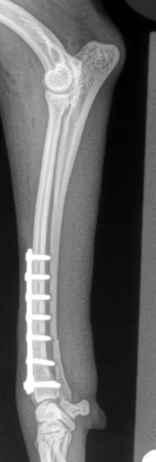
Distal radius-ulna fractures are very common in small/toy breed dogs and the Italian Greyhound as a result of jump down trauma. These frequently simple, transverse, two-piece fractures are commonly managed by open, anatomic, radius bone reduction and plate application in compression.
Recovery and Prognosis
Healing may be quicker and easier with these patients and situations:
- Young animals
- Calm and quiet
- Healthy
- Small, medium, or large breeds
- One limb injury
- Closed or transverse fractures
- Minor surgical trauma
- Under close supervision
- Restricted activity
Healing may be slower or difficult with these patients and situations:
- Older animals
- Bouncy and active
- Underlying medical conditions
- Toy or giant breeds
- Multiple fractures or internal organ injuries
- Open or comminuted fractures
- Moderate surgical trauma
- Inadequate owner supervision
- Unrestricted activity
For sufficient healing, fractures in puppies generally require a minimum of four to six weeks, and older animals require approximately eight to twelve weeks. After the fracture heals, the pet may gradually return to physical activity, and the surgeon will provide care and recovery instructions to aid healing.
We do. When a pet is injured, owners and surgeons are on the same page – accurately determine what’s wrong, decide the best course of action, and administer treatment as safely and efficiently as possible. We’ve successfully cared for several hundred animals with fractured bones and helped them return to living active, happy lives.
Fractures aren’t usually life-threatening. Once healed, most pets enjoy an active and normal life. Concurrent damage to internal organs or the spine may slow the healing process and have a less desirable prognosis.
On average, broken bones gradually heal in two to four months. Recovery time is variable for each dog based on:
- Their age
- Fracture type and severity
- Underlying medical conditions
- Coexisting internal injuries besides the fracture
- Your adherence and willingness to follow postoperative instructions
Caring for your dog shouldn’t be difficult, but it could be challenging because you’ll need to take precautions to protect the healing bone despite their wanting to run and jump. Dogs don’t understand how to take it easy, so too much activity may be detrimental, especially during the early phases of healing.
To make home care manageable, we’ll provide instructions to aid recovery, including:
- Bandage care
- Confinement
- Activity restriction
- Helping them stand and walk
- Using cold-packs to ease inflammation and pain
- Directions for passive range of motion (PROM) stretches, massage, and a gradual return to activity
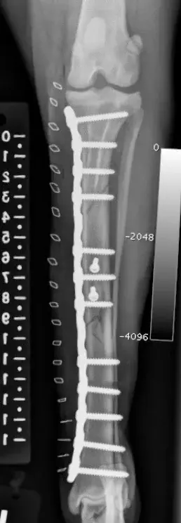
This large segmental tibia-fibula fracture in an Irish Wolfhound was managed by interfragmentary screws and a large medial bone plate applied in a neutralization fashion.
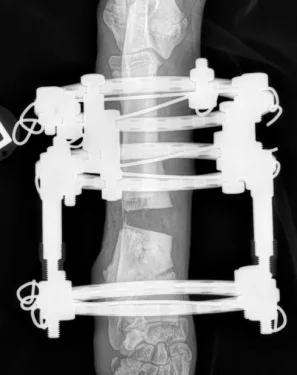
This image demonstrates the use of circular ring external skeletal fixation for gradual and incremental correction of limb alignment and limb lengthening of the radius-ulna in a dog. This device uses linear “motors” to distract and move the bones daily by use of a simple wrench. Limb lengthening may be achieved at a rate of approximately 1 mm per day.
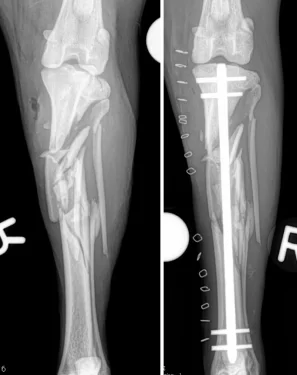
This highly comminuted tibia-fibula fracture in a Border Collie was managed with a Biomedtrix I-Loc interlocking nail (ILN). Many ILN applications in the tibia of dog’s may be managed without opening of the fracture site and disruption to the fracture hematoma and local blood supply. These minimally-invasive techniques may help bones heal faster and with less complications.