The ability to view bones and joints in 3 dimensions has clear advantages when compared to imaging with standard 2D radiographs, but even better still, the data from a CT scan can be used to create life-sized anatomic bone/joint models as well as custom, patient-specific, bone cutting and reduction guides.
Advanced CT Imaging and 3D Modeling
Normal anatomy can be complex but certain orthopedic conditions pose particular diagnostic challenges due to anatomic abnormalities, small regions of interest or superimposition of body parts.
Advanced cross-sectional imaging by computed tomography (CT) is commonly performed for assessment of orthopedic conditions in dogs and cats. Traditionally, these images are viewed using special software which allows us to look at different planes (or slices in different directions) and for the creation of a virtual model of the bones and tissues that can be manipulated on a computer screen, allowing for viewing from different angles. These 2D and 3D images can also be used to virtually “rehearse” surgical procedures and plan for implants if needed.
Patient-specific implants including bone plates or joint replacement components can be created using 3D metal printing technology. Together this approach allows for the customization of surgical procedures and patient-specific planning.
3D planning and printing has a wide variety of applications in veterinary orthopedics. In the simplest form, 3D models of normal and abnormal anatomy can be created to use in educating pet owners on conditions and treatment options. 3D printed bone models can be used to rehearse procedures prior to surgery – reviewing steps and optimizing implant placement. They can be sterilized and brought into the OR for improved spatial understanding of anatomy.
Patient-specific surgical planning is increasingly being used to treat bone deformities of front and hind limbs, high-grade patella luxation, bone/joint trauma, and for complex joint replacement. In these cases, challenging anatomic abnormalities are overcome by combining virtual surgical planning with printing to allow for the creation of patient-specific instrumentation and guides. In surgery, 3D printed guides are applied precisely to the bone using the individual patients’ anatomic landmarks where they are used to direct osteotomies, maintain accurate reduction and guide implant placement.
What Are the Benefits of Patient-Specific Surgical Planning?
3D printed models can be used to help pet owners better understand complex orthopedic conditions and the surgical options for managing them. The use of patient-specific surgical planning and 3D printing helps to improve the accuracy and precision of surgery, reduces intraoperative decision making, allows for precise implant placement and decreases surgical time – all of which may help to improve patient outcome, reduce infection risk and speed recovery.
What Conditions May Be Treated with Patient-Specific Planning and 3D Printing (3DP)?
A CT scan may be recommended in the workup of any number of conditions however not all of these will require 3D printing or planning. The most common conditions managed with 3DP include bone deformities of the front and back legs and high-grade patella luxation. These conditions generally have predictable (though to varying degree) abnormalities of the associated bones/joints, the development of surgical plans and guides are well established and the outcomes are proven. Other conditions that may benefit from 3D printing include complicated joint replacement, developmental orthopedic diseases, trauma (including complex fractures of the long bones, pelvis, skull or spine) and for use with minimally-invasive bone and joint surgeries.
What Is the Process for Creating a Patient-Specific Surgical Plan?
- Evaluation – as with all conditions, the process starts with a thorough examination performed by one of our surgeons. In many cases, sedated examination and radiographs will be recommended to obtain anatomic measurements and to use for surgical planning and follow-up evaluations.
- CT – CT scans are performed offsite, on an outpatient basis through the Veterinary Neurological Center. Once it is determined that a CT scan is necessary, a referral will be submitted. You will then be able to schedule imaging directly through VNC.
- Imaging Review – Once the CT scan is complete, we will review the study and discuss the findings with you. If patient-specific planning is indicated, we will review your pet’s condition and imaging with colleagues who provide surgical planning and printing support.
- Surgical plan review – Once the surgical plan is complete, we will review this with our planning team before presenting it to you. Once the surgical plan is confirmed, a build date and expected delivery date for models and guides will be established.
- Delivery of models and guides – 3D printed models and guides will be delivered to ACOSM prior to the planned surgery date. At times, surgery may not be scheduled until after the items are received to allow for preparation. Models are typically received within 4-6 weeks of the CT scan however it is sometimes necessary to coordinate the timing of imaging and model creation more closely in young, growing patients in order to maintain accuracy of planning. Once the models are received, your surgeon will verify the surgical plan, rehearse the procedure if necessary and prepare surgical implants.
- Surgery is performed.
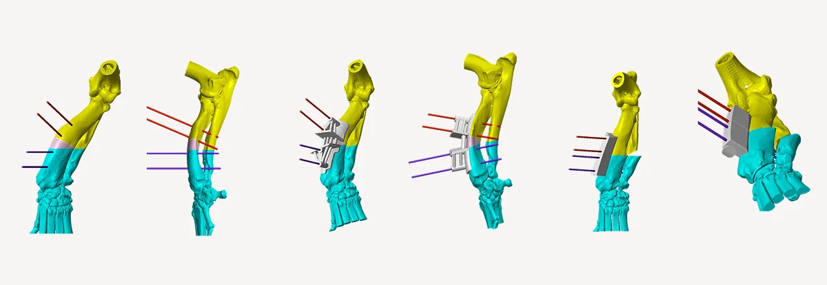
Below is an example of a common forelimb angular deformity as a consequence of premature closure of the distal ulna physis. Growth plate injury and resulting asynchronous growth between the radius and ulna bones leads to bowing of the radius, valgus deviation of the paw and elbow subluxation. Corrective osteotomy treatment can be very challenging especially in chondrodystrophic breeds with already very short limbs.
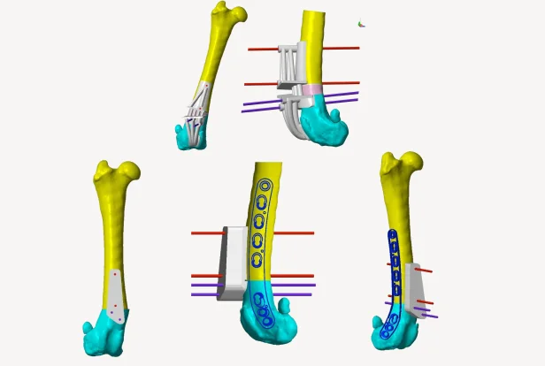
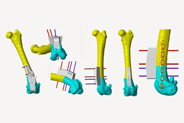
This next example is a Labrador Retriever with a high grade medial patella luxation and an underlying conformation issue known as distal femoral varus – or bowed legs. Treatment of the patella luxation, in this case, requires improving the alignment of the quadriceps/patella mechanism by straightening of the femur bone n a procedure known as a Distal Femoral Osteotomy (DFO).
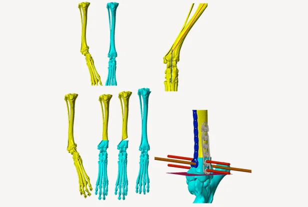
This last example is a premature, partial closure of the distal tibial growth plate (physis). As the outer half of the tibia cotinued to grow and develop, the foot began to twist and bend inward. Corrective osteotomy is planned by either a closing or opening wedge osteotomy with orthogonal, double bone plating.